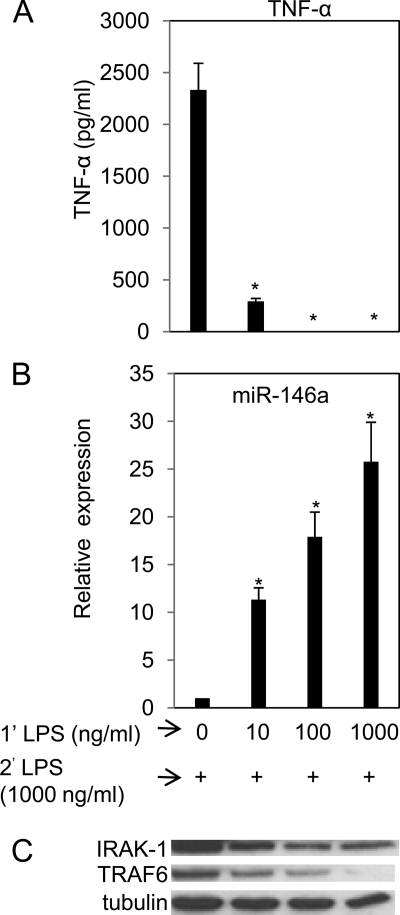
FIGURE 3.
Higher priming dose of LPS induced higher miR-146a levels and more efficient suppression of TNF-α production in subsequent LPS challenge. THP-1 cells were primed with 0, 10, 100, or 1000 ng/ml LPS continuously for 18 h, washed twice with PBS, and challenged with 1000 ng/ml LPS. Supernatants and cell pellets were collected 5 h later as described under “Experimental Procedures” for TNF-α protein determined by ELISA (A), miR-146a expression analysis in total RNA (B). Data points and error bars represent mean ± S.D. of three independent experiments. *, p < 0.01 compared with untreated cells. Western blot analysis for IRAK-1, TRAF6, and tubulin in the cell lysates collected 2 h after LPS challenge (C).

