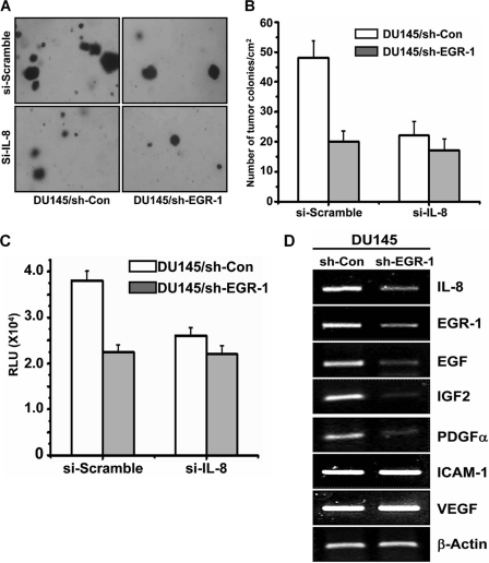
FIGURE 5.
Knockdown of Egr-1 inhibited IL-8-mediated tumor colony formation and invasion. A and B, tumor colony formation was performed with DU145/si-con and DU145/si-Egr1 cells that were further transfected with siRNA silencing IL-8 or scramble RNA. The cells were plated in a 6-well plate (1 × 10⁴ cells/well) and cultured for 14 days. Cell colonies were visualized under a microscope (A), and the number was counted and presented as number/cm2 (B). C, for the chemoinvasion assay, the transfected cells were plated onto Matrigel-coated wells and cultured for 48 h. By the end, the cells in the lower chamber of the transwell were collected and measured by CyQUANT@GR Dye (QCMTM 24-Well Cell Invasion Assay kit) per the manufacturer's instruction. The data are presented as relative luminescence units and the mean of three samples in each experiment. D, gene expression profile of other growth factors and invasion proteins measured by RT-PCR.

