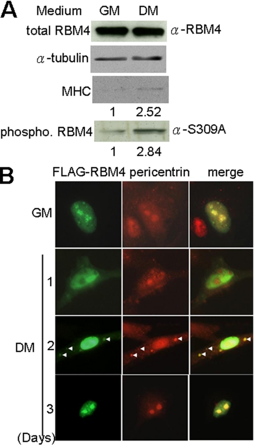
FIGURE 1.
Phosphorylation and subcellular localization of RBM4 during myoblast differentiation. A, immunoblotting of cell lysates prepared from C2C12 cells that were cultured in growth (GM) or differentiation medium (DM) for 2 days. Relative level of myosin heavy chain (MHC) protein expression and RBM4 phosphorylation in GM versus DM is indicated below the gels. B, immunofluorescence of C2C12 cells that transiently expressed FLAG-RBM4 and were cultured in GM or DM for 1–3 days.

