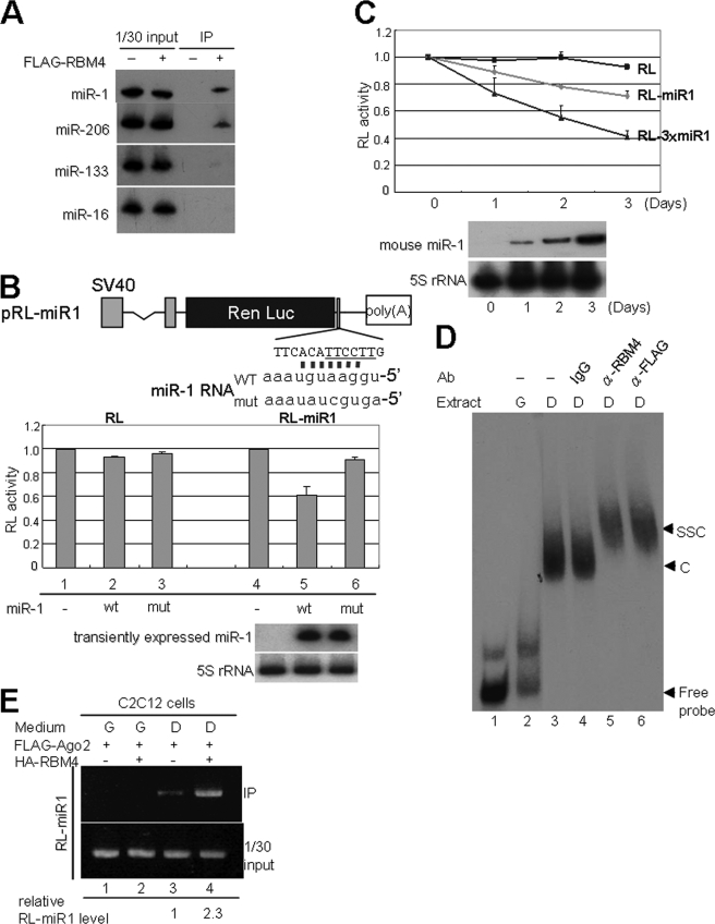
FIGURE 4.
RBM4 selectively interacts with miRNAs and promotes Ago2 association with miRNA targets. A, FLAG-RBM4 was immunoprecipitated (IP) from differentiated C2C12 cells. Coprecipitates were analyzed by Northern blotting. B, diagram shows the pRL-miR1 reporter containing a synthetic miR-1 targeting sequence; the CU-rich sequence is underlined. The wild-type (WT) and mutant (mut) miR-1 RNAs are also depicted. The pRL or pRL-miR1 reporter was cotransfected with miR-1 expression vector in HEK293 cells. The reporter assay was essentially as in Fig. 3. Expression of miR-1 RNA was detected by Northern blotting in both B and C. C, reporter assay using pRL-miR1 or pRL-3×miR1 was performed in C2C12 cells that were cultured in DM for 0–3 days. D, EMSA was performed using 32P-labeled RL-3×miR1 3′-UTR RNA as probe and C2C12 cell cytoplasmic extract containing FLAG-Ago2. C and SSC represent RNA-protein complex and supershifted complex, respectively. E, immunoprecipitation-RT-PCR was as similar to Fig. 3D (lanes 1–3), except that transfection was performed in C2C12 cells. Relative amount of coprecipitated RL-miR1 RNA is indicated. G, GM; D, DM.

