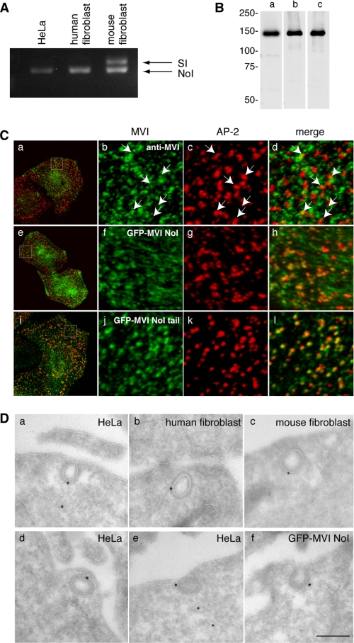
FIGURE 1.
The myosin VI NoI isoform localizes to CCS at the plasma membrane. A, PCR analysis of myosin VI isoforms expressed in nonpolarized cells. HeLa cells and human wild type fibroblasts express the myosin VI NoI isoform, whereas mouse wild type fibroblasts express both the NoI and the SI isoforms. B, Western blot analysis of HeLa whole cell lysates using different myosin VI antibodies: an antibody against the C-terminal tail (lane a), an antibody against the whole tail (lane b), and the C-terminal tail of myosin VI (lane c). C, co-localization of myosin VI (a–d), GFP-myosin VI NoI (e–h), and GFP-myosin VI NoI tail (i–l) with AP-2. The boxes in the merged image in a, e, and i indicate the areas enlarged in the adjacent panels b–d, f–h, and j–l, respectively. Arrows indicate examples of co-localization. D, to visualize myosin VI localization at the ultrastructural level in unpolarized cells, cryosections of HeLa cells and human and mouse wild type fibroblasts were immunogold-labeled with a commercial antibody to the C-terminal region of myosin VI (a–c), an antibody to the whole tail (d), or an antibody the C-terminal tail of myosin VI (e). Cryosections of HeLa cells expressing GFP-myosin VI NoI were labeled with an antibody against GFP (f). Bars: a, 200 nm; b, 200 nm; c, 300 nm; d, 200 nm; e, 200 nm; f, 200 nm.

