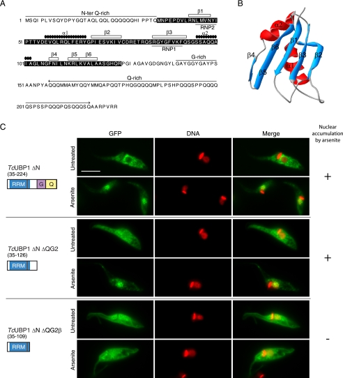
FIGURE 3.
TcUBP1 RRM mediates arsenite-induced nuclear accumulation. A, primary and secondary structure of TcUBP1. The RRM is shown boxed in black. Secondary structure elements, Gln and Gly-rich regions, and RNP1 and RNP2 are indicated. B, ribbon diagram of amino acids 41 to 120 of TcUBP1 comprising the RRM, based on NMR structural data. Secondary structure elements are indicated. β5 and β6 strands are shown together. C, localization of different TcUBP1 deletion mutants expressed as GFP fusion proteins in untreated and arsenite-treated (2 mm, 4 h) transfected parasites. Mutant protein names and a scheme of the domains fused to GFP are shown on the left side of the respective images. Numbers in parentheses indicate the amino acid residues fused to GFP. DNA was stained with DAPI, and is shown in red for better contrast. The column on the right summarizes if the fusion protein accumulates (+) or not (−) in the nucleus of the parasites under arsenite treatment. Scale bar, 5 μm.

