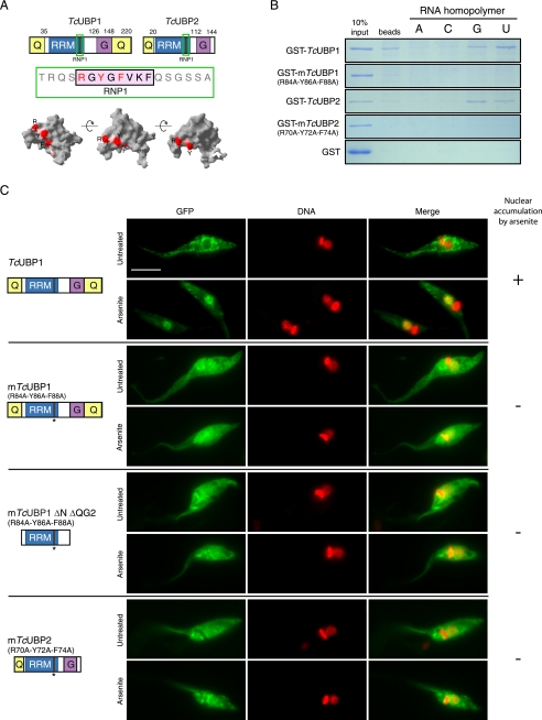
FIGURE 4.
RNP1 mutations affect RNA binding and arsenite-induced nuclear accumulation. A, TcUBP1 and TcUBP2 protein schemes showing the RRM and accessory domains. The fragment containing the RNP1 peptide is shown in green and the amino acids mutated to alanine in mTcUBP1, mTcUBP1 ΔN ΔQG2, and mTcUBP2 are shown in red (R, Y, F). Different views of TcUBP1 RRM based on NMR spectroscopy data showing the RNP1 peptide with the mutated amino acids in red are indicated. Structure analysis was performed using Swiss-Pdb Viewer version 3.7 using TcUBP1 RRM as input (Protein Data Bank code 1U6F). B, RNA homopolymer binding specificity of TcUBP1, mTcUBP1, TcUBP2, and mTcUBP2. Proteins were synthesized as recombinant GST fusions and incubated with dihydrazide-agarose beads alone (beads) or cross-linked to homoribopolymers (A, C, G, or U). After washing and elution, SDS-PAGE was performed, and the gel was stained with Coomassie Brilliant Blue R-250. C, localization of TcUBP1, mTcUBP1, mTcUBP1 ΔN ΔQG2, and mTcUBP2 as GFP fusion proteins in untreated and arsenite-treated (2 mm, 4 h) transfected parasites. Protein names and a scheme of the protein fused to GFP are shown on the left side of the respective images. Numbers in parentheses indicate the amino acid residues mutated to alanine (parental numeration). TcUBP1 is shown for comparison with mutants. DNA was stained with DAPI, and is shown in red for better contrast. The column on the right summarizes if the fusion protein accumulates (+) or not (−) in the nucleus of the parasites under arsenite treatment. Scale bar, 5 μm.

