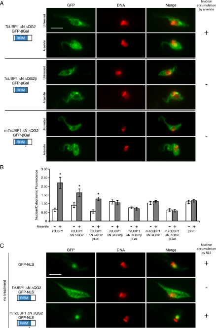
FIGURE 5.
Nuclear import and export of TcUBP1 RRM. A, behavior of TcUBP1 mutants fused to GFP-βGal. The localization of TcUBP1 ΔN ΔQG2, TcUBP1 ΔN ΔQG2β, and mTcUBP1 ΔN ΔQG2 fused to GFP-βGal is shown in untreated or arsenite-treated (2 mm, 4 h) parasites. The column on the right summarizes if the fusion protein accumulates (+) or not (−) in the nucleus of the parasites under arsenite treatment. B, nuclear/cytoplasmic fluorescence ratio obtained from parasites from different transfection experiments, in the two tested conditions. The ratios of TcUBP1 and GFP are the same as Fig. 1B. Asterisks (*) indicate statistically significant difference (p < 0.00001) within the same population. C, the localization of GFP-NLS, TcUBP1 ΔN ΔQG2-GFP-NLS, and mTcUBP1 ΔN ΔQG2-GFP-NLS is shown in untreated parasites. DNA was stained with DAPI, and is shown in red for better contrast. The column on the right summarizes if the fusion accumulates (+) or not (−) in the nucleus due to the NLS. Scale bar, 5 μm.

