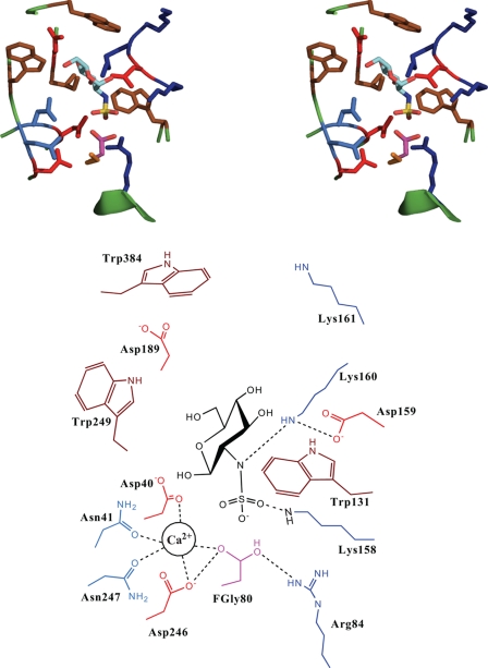
FIGURE 3.
Theoretical structural model of N-sulfamidase-GlcNS substrate. Shown on top is the stereo view of the active site of N-sulfamidase with the docked GlcNS substrate. The side chains of the key residues are shown and colored as follows: Arg and Lys, blue; Asp and Glu, red; Trp, Leu, and Ile, brown; Asn and Gln, light blue; and FGly, purple. The Glc-NS substrate is colored by atom as follows: C, cyan; O, red; N, blue; and S, yellow. Shown at the bottom is the schematic of the active site with the key amino acids labeled for clarity. Also shown in the schematic is the location of the divalent Ca2+ ion and its interactions with the active site and the substrate.

