
Fig. 1.
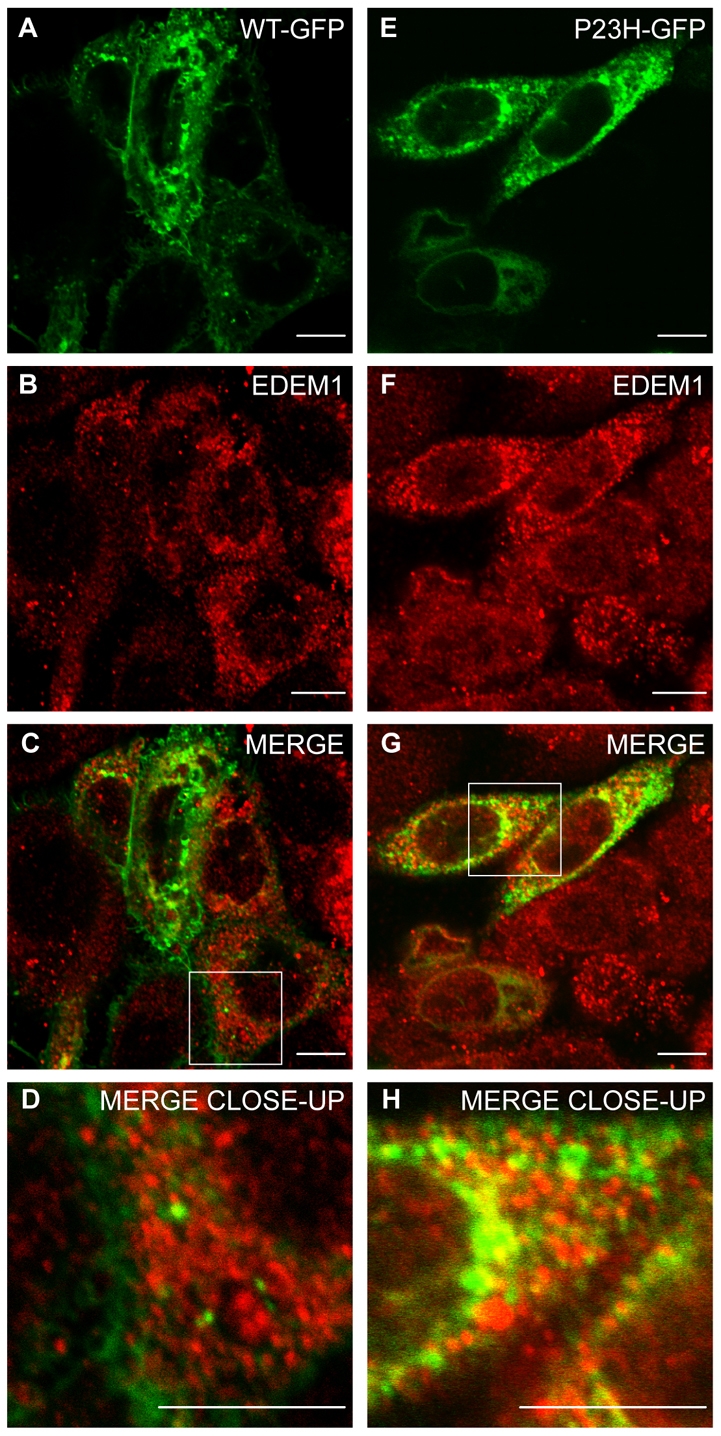
Endogenous EDEM1 partially colocalises with P23H but not WT rod opsin. SK-N-SH cells were transfected with WT rod opsin-GFP (WT-GFP) (A-D, green) or P23H rod opsin-GFP (P23H GFP) (E-H, green). 24 hours after transfection, the cells were fixed and stained with antibodies against EDEM1 (red). Cells were analysed by confocal microscopy. Areas enclosed by white boxes in C and G are enlarged in D and H to show detail of rod opsin-GFP and EDEM1 staining. Scale bars: 10 μm.