Fig. 1.
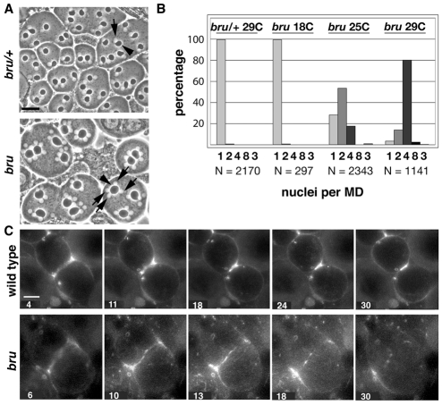
Contractile ring constriction is inhibited in bru mutant spermatocytes. (A) Phase-contrast images of early spermatids in bruZ3358/CyO (bru/+) and bruZ3358/Df(2L)pr2b (bru) testes from males raised at 29°C. CyO is a balancer chromosome that provides wild-type bru function, and Df(2L)pr2b is a deficiency that deletes bru. Note that testis squash preparations typically disrupt the plasma membrane separation between early spermatids. Arrowheads indicate MDs and arrows indicate nuclei. Scale bar: 20 μm. (B) Percentages of early spermatids with 1, 2, 4, 8 or 3 nuclei per MD from bruZ3358/+ and bruZ3358/Df(2L)pr2b males raised at the indicated temperatures. N, total number of spermatids counted. (C) Behavior of contractile rings labeled with RLC-GFP in wild-type and bru mutant spermatocytes. Selected frames from supplementary material Movies 1 and 2 showing wild-type (+/+) and bruZ3358/Df(2L)pr2b (bru) spermatocytes raised at 25°C. Spermatocytes were imaged for 30 minutes starting from late anaphase. The numbers at the bottom of each frame indicate time elapsed from the beginning of imaging. In the wild type, the RLC-GFP ring constricts fully. By contrast, the RLC-GFP ring in the bru mutant spermatocyte undergoes only slight constriction and eventually breaks down. Scale bar:10 μm.

