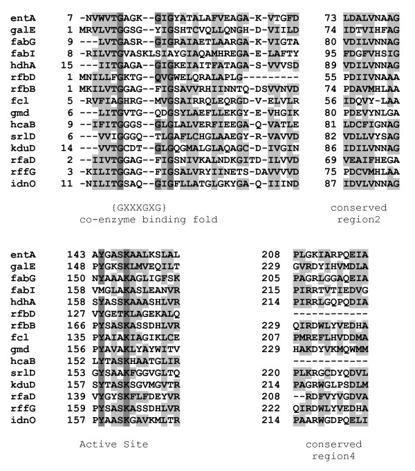
Figure 2.
Alignment of E. coli SDR family members. The enzymes of the family members are listed in Table 1. Four conserved regions of the proteins are shown. The protein sequences were aligned with ClustalW 2.0.11. Identical residues are highlighted in dark grey while conserved and semi-conserved residues are highlighted in light grey.

