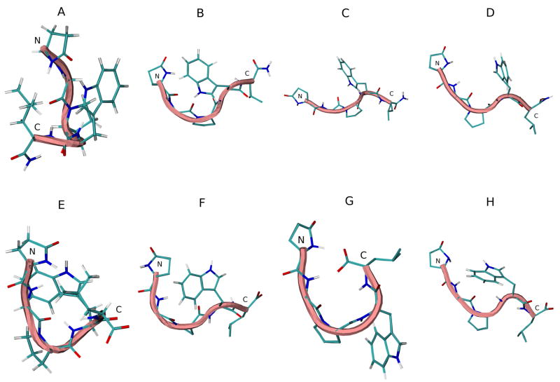
Figure 7.
Initial energy minimized structure and middle structures of the most populated cluster of G17(1-5)-NH2 (A–D) and G17(1-5) (E–F) in various solvents: A and E, initial structure; B and F, H2O; C and G, DMSO; D and H, TFE. Backbone structure is color coded: blue, representing 310 helix; pink, random meander structure; cyan, representing β-turn structure. N and C indicate the N- and C-terminus, respectively.

