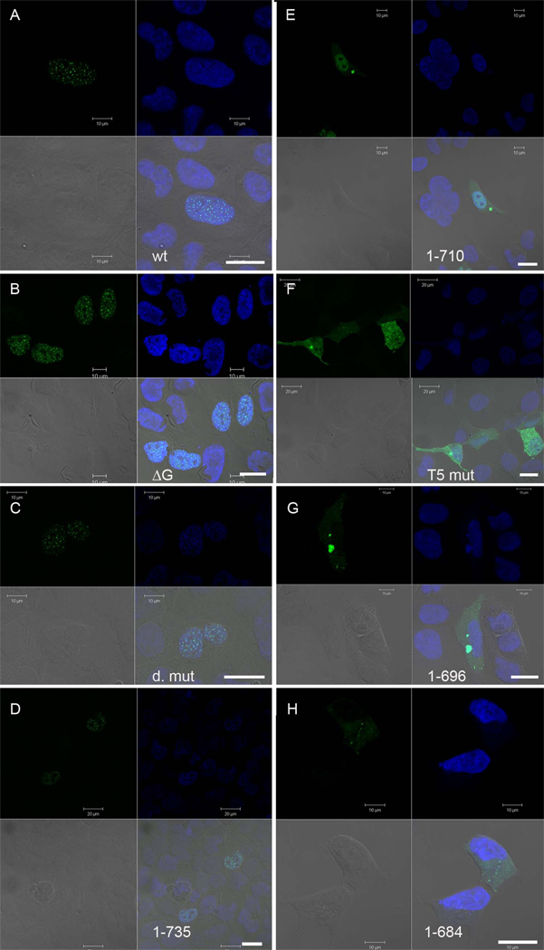
Figure 2. Identification of a novel NLS, and a sequence element directing TFIP11 to distinct nuclear speckles using confocal microscopy.
Direct fluorescent images of TFIP11 transfected cells using either the wild-type TFIP11-C1 construct (panel A), or mutant constructs as identified in figure 1: TFIP11ΔG-patch (panel B); double mutant (panel C); TFIP111–735 (panel D); TFIP111–710 (panel E); V701KDKFN→T701TTTT (panel F); TFIP111–696 (panel G); and TFIP111–684 (panel H). Cells counterstained with DAPI, and the merged image is seen in the lower right quadrant of each panel. Also seen in the lower right quadrant is a scale bar (thick white line) for 20µm. Additional scale bars for either 10µm or 20µm, added in at the time of imaging, can be seen in each panel.

