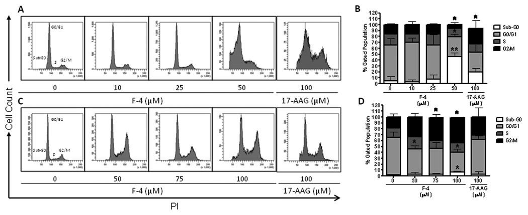
Figure Six. Cell cycle effects of F-4 in prostate cancer cells.
Representative histograms are shown depicting the distribution of cell cycle as assessed by PI staining following treatment of LNCaP (panel A) and PC-3 (panel C) cells with F-4 or 17-AAG for 72h. Bar graphs depicting the cell cycle distribution after treatment from three independent experiments are shown with statistical analysis for LNCaP and PC-3 (panels B and D, respectively). Asterisk(s) *, ** indicates significant P value <0.05 and <0.01, respectively, by two-tailed t-test compared to vehicle-treated control.

