
Fig. 3.
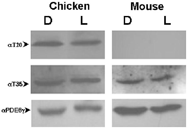
PDE6H phosphorylation in chicken and murine retina.
Immunoblot of normalized dark- and light-adapted chicken and murine retinal extracts probed with rabbit anti-phosphoT20 {TTGDAPTGPT(pT)PR} antibody recognizing the phosphorylated T20 residue of PDE6H or rabbit anti-pT35 antibody recognizing the phosphorylated T35 residue of PDE6γ. Phosphorylated T20 was not detected in mouse retinal extract. Presence of PDE6γ in each sample was confirmed by stripping and re-staining the filter with rabbit anti-native PDE6γ antibody. D, dark-adapted overnight; L, light-adapted to 1.67×10⁻¹ W/cm2 for sixteen hours.