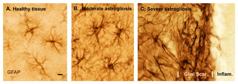
Figure 1. Photomicrographs of astrocytes in healthy tissue and of different gradations of reactive astrogliosis and glial scar formation after tissue insults of different types and different severity.
(A-C) Immunohistochemical staining of glial fibrillary protein (GFAP) in wild-type mice. Note that GFAP staining demonstrates the main stem processes and general appearance of the astrocytes, but does not reveal all of the fine branches and ramifications. (A) Appearance of ‘normal’ astrocytes in healthy cerebral cortex of an untreated mouse. Note that the territories of astrocyte processes do not overlap. (B) Moderately reactive astrogliosis in mouse cerebral cortex in response to intracerebral injection of the bacterial antigen, lipopolysaccharide (LPS). Note that the territories of moderately reactive astrocyte processes also do not overlap. (C) Severely reactive astrogliosis and glial scar formation adjacent to a region of severe traumatic injury and inflammation (Infam.) in the cerebral cortex. Note the extensive overlap and inter-digitations of processes of severely reactive and scar-forming astrocytes. Scale bar = 8μm.

