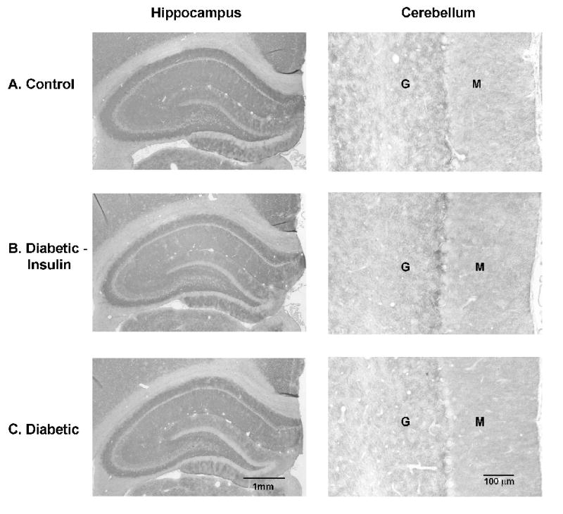
Figure 5. GLT-1.

Immunohistochemistry of astrocytic GLT-1 in the hippocampus and cerebellum after 4 weeks of diabetes. No obvious changes were detectable in the intensity of immunoreactivity among the different treatment groups. (Control: n = 8; Diabetic + Insulin: n = 8; Diabetic/no insulin: n = 8). Similar findings were seen after 8 weeks of diabetes. Representative sections are shown. M: molecular layer; G: granular layer.
