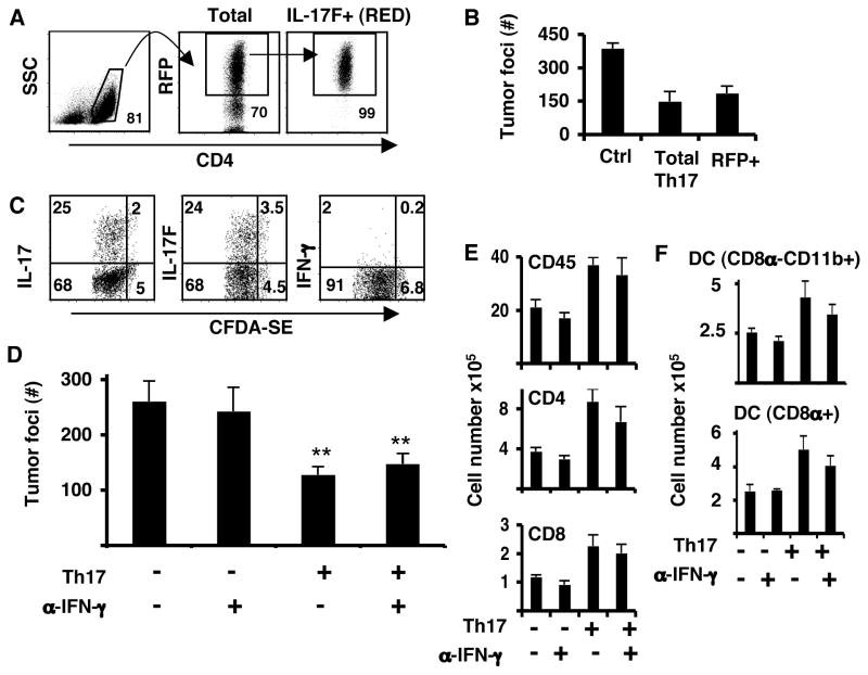
Figure 4. Th17 cells maintain their cytokine expression in tumor-bearing mice.
Purified CD4+ T cells from OT-II.IL-17F-RFP reporter mice were cultured in Th17 conditions. On day 4, CD4+RFP+ cells were sorted and injected into C57BL/6 mice bearing 5-day established pulmonary B16-OVA tumors. A. Sorting strategy for RFP+, IL-17F producing cells. B. Tumor foci from lungs of C57BL/6 mice that had B16-OVA melanoma and received either no treatment (C), unsorted Th17 (total Th17) or RFP+ sorted (IL17F+ RED). C. Purified CD4+ T cells from Rag1−/− OT-II mice were cultured in Th17 conditions and on day 4, cells were labeled with CFDA-SE and transferred into C57BL/6 mice bearing 5-day established pulmonary B16-OVA tumors. LLN from these mice were analyzed for proliferation and IL-17, IL-17F and IFN-γ production on day 4 after transfer. D. Mice were inoculated with B16-OVA tumor and received on the same day Th17 (Th17) cells i.v or no T cells. A set of mice from each group was treated with anti-IFN-γ blocking antibodies (α-IFN-γ) on day -1 and every other day until day 14 after tumor challenge. Shown are the tumor colonies present in the lung lobes of each group of mice (n=4, avg. +/− s.d). (**P<0.01). E. Total cell numbers from leukocyte lung fractions. Number were calculated from the percentages of live cells gated on CD45.2 (n=4, avg. +/− s.d). F. Number of myeloid cell populations from leukocyte lung fraction was calculated from the percentage of live cells gated on CD45.2 (n=4, avg. +/− s.d.).

