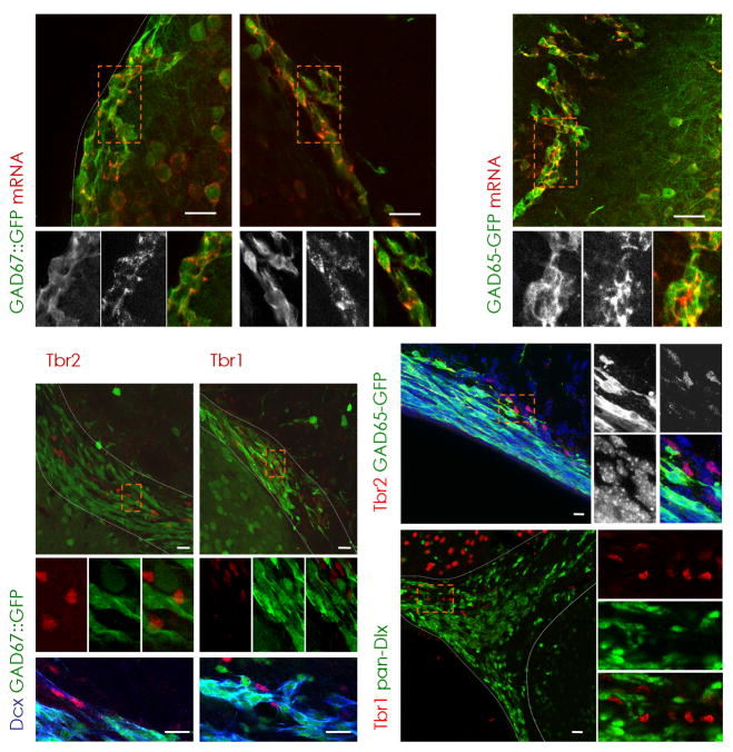
Figure 3. Presence of Tbr2 and Tbr1 defines a non-GABAergic subpopulation of neuroblasts.
(a–c) Virtually all GFP-positive cells in the SEZ (a,c) and RMS (b) are colocalizing with GAD67 (a,b) or GAD65 (c) mRNA in the respective GAD67::GFP and GAD65-GFP mouse lines as indicated in the panels (see Methods). Notably, Tbr2 (d,h) and Tbr1 (e) are absent in GAD65- (h) or GAD67- (d–g) driven GFP-positive cells, but are immunoreactive for the neuroblast marker Dcx in the dorsal SEZ (Tbr2, f) and RMS (Tbr1, g). (i) Consistently, Dlx transcription factors (pan-Dlx, green) are not coexpressed with Tbr1 (red). (a–e,h,i) Boxed areas (orange) are shown at higher magnification. Nuclei are visualized with DAPI (h).
dSEZ = dorsal subependymal zone, LV = lateral ventricle, RMS = rostral migratory stream, SEZ = subependymal zone, Str = Striatum, WM = white matter, latSEZ = lateral subependymal zone, scale bars: 20 μm.

