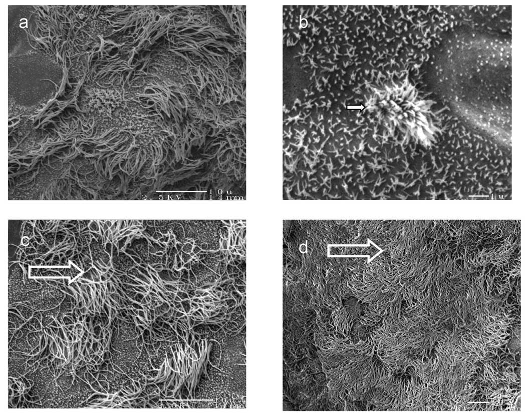
Fig. 3.
Scanning electron micrographs (SEM) of non-cryopreserved ovine tracheal epithelial cell cultured in air-liquid interface for 2 weeks. Samples were sputter-coated with Pt/Pd. (a) ciliogenesis was captured at several stages (bar=10 µm), (b) magnified SEM picture showing an active stage of ciliogenesis such as the budding cilia (arrow) among microvilli (star) (bar=1 µm), (c) mature cilia reached a length of approximately 6–7 µm (bar=10 µm), (d) over 50% of the cultured cell surface was covered with mature cilia (bar=10 µm).

