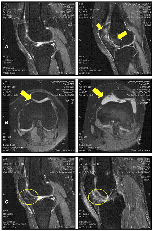
Figure 1.
A. Sagittal MRIs of the knee. The left MRI is of a knee without any BMLs. The right MRI is of a knee with a large BML in the lateral patella and lateral trochlea (yellow arrows).
B. Axial MRIs of the knee. The left MRI is of a knee with a physiologic effusion. The right MRI is of a knee with a large joint effusion.
C. Sagittal MRIs of the knee. The left MRI is of a knee without any infrapatellar synovitis. The right MRI is of a knee with a large amount of synovitis in the infrapatellar fat pad (yellow circles).

