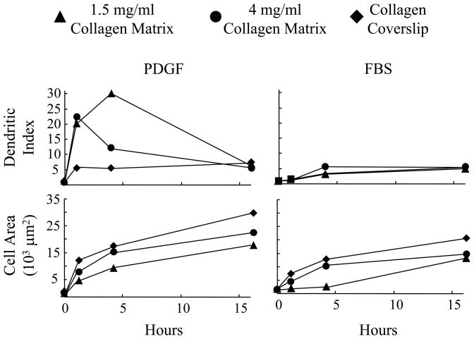
Figure 3. Morphometric analysis of fibroblast spreading in PDGF and serum media on 1.5 mg/ml and 4 mg/ml collagen matrices and collagen-coated coverslips.
Fibroblasts were cultured 1 hr, 4 hr and 16 hr on 1.5 mg/ml and 4 mg/ml collagen matrices and collagen-coated coverslips in PDGF and serum media. At the end of incubations, cells were fixed and stained for actin and evaluated by morphometric analysis. Marked differences between PDGF and serum media in formation of dendritic extensions and time of cell spreading occurred for cells on 1.5 mg/ml collagen matrices. These differences were less evident with 4 mg/ml collagen matrices and not detected with collagen-coated coverslips.

