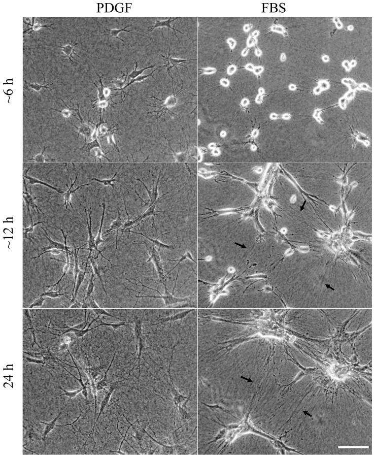
Figure 4. Cell migration in PDGF medium vs. clustering in serum medium.
Phase-contrast images from videos of fibroblasts interacting with 1.5 mg/ml collagen matrices in PDGF (Supplemental Video 1) and serum media (Supplemental Video 2) with the same field shown after ~6hr, ~12 hr, and 24 hr. Alignment of collagen fibrils between cells (arrows) becomes increasingly apparent in serum but not PDGF medium. Cells moved as individuals in PDGF medium but contracted into clusters in serum medium. Scale bar, 100 μm.

