
Figure 7.
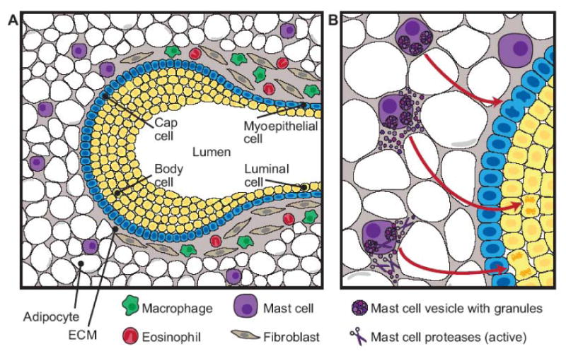
Diagram of innate immune cells that contribute to the stromal effects on mammary gland branching morphogenesis. A) The diagram depicts the stromal locations of eosinophils, macrophages, and mast cells relative to a terminal end bud (TEB). The TEB consists of a single layer of cap cells and multiple layers of body cells, which will eventually make up the luminal cells of the mature duct surrounded by a myoepithelial layer. The area surrounding the neck of the TEB consists of a collagen-rich stroma including fibroblasts, macrophages, and eosinophils. At the invading edge of the TEB, a thin basement membrane separates the advancing TEB from the adipocyte-rich stroma. Mast cells are found around the TEB in the stromal compartment, often just ahead of the advancing epithelium. B) Depiction of the effect of mast cell activation on mammary gland development. Mast cell degranulation and subsequent release of active proteases stimulate epithelial cell proliferation, promoting TEB formation and duct branching in the pubertal gland.