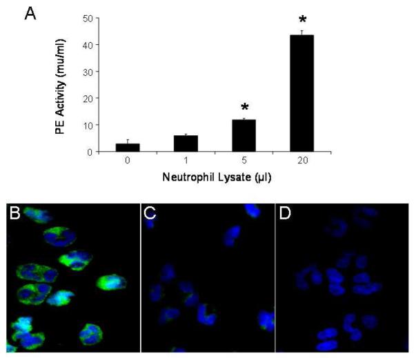
Figure 1.
Indicated amounts of human neutrophil lysate (106 cells) were assayed for PE enzymatic activity (expressed as mU/ml). Results are presented as mean ± SEM (A) and compared using the two group t test. Increased PE activity was observed with greater amounts of neutrophil lysate (* p < 0.05 compared with 0μl lysate, n = 3 per group). Results are representative of several experiments. Cytospins of human peripheral blood neutrophils on glass slides were incubated with anti-PE antibody (B), anti-PE antibody pre-adsorbed with PE (C) or pre-immune rabbit antibody (D), followed by FITC-labeled goat anti-rabbit IgG, and examined by immunofluorescence microscopy. PE in human neutrophils was located in the cytoplasm in a granular pattern (B).

