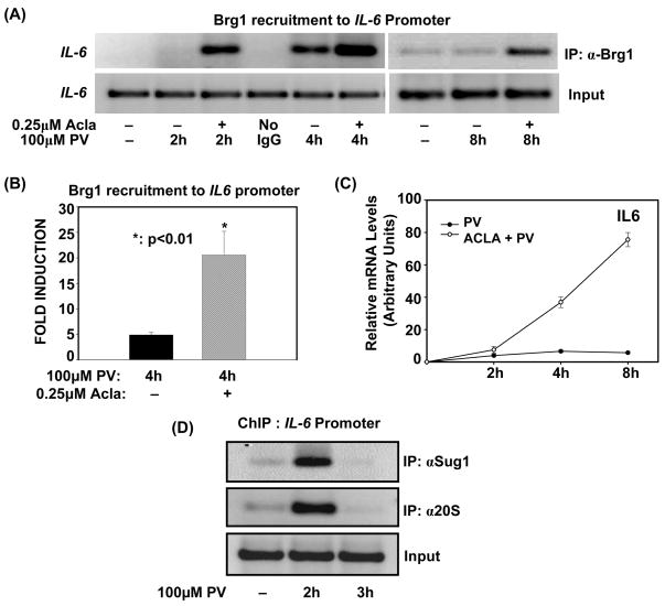
Figure 1. Proteasome activity limits Brg1-promoter association with kinetics consistent with transcriptional induction and shut-off.
(A). ILU-18 cells were either untreated or treated with PV (100μM) for 2, 4 and 8 hours, with (+) or without (-) pretreatment with Acla (0.25μM) for 2 hours. ChIP assay employing anti-Brg1 was performed using immunoprecipitated DNA amplified with promoter-specific primers for IL-6
(B). ChIP assays were performed on ILU-18 cells treated with PV (100μM) for 4 hours, with (+) or without (−) pretreatment with Acla (0.25μM) for 2 hours using Brg1 antibody for the immunoprecipitation. The immunoprecipitated chromatin was submitted to qPCR analysis using primers amplifying the promoter region of IL-6. Fold induction represents anti-Brg1 ChIP relative to control ChIP (immunoprecipitated DNA from untreated cells). Statistically significant differences are denoted by * (p value < 0.05)
(C). ILU-18 cells were stimulated with PV (100μM), with (○) or without (●) pretreatment with Acla (0.25μM) for 2 hours. Total RNA was extracted at indicated times and analyzed by RT-qPCR for mRNA expression levels of IL-6. The relative mRNA levels were determined from two independent experiments and are presented as mean ± standard error.
(D). ChIP assays employing antibodies recognizing Sug1 and 20S were performed using immunoprecipitated DNA amplified with promoter-specific primers for IL-6. Chromatin was obtained from ILU-18 cells which were stimulated with PV (100μM) for 0, 2, and 3 hours.

