Figure 2.
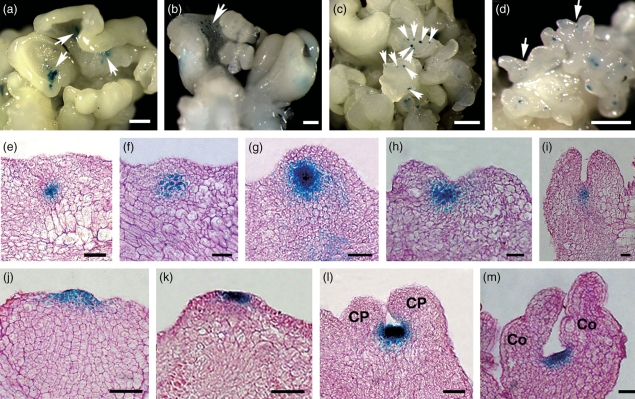
Patterns of pWUS::GUS and pCLV3::GUS expression during somatic embryogenesis by light microscopy. (a) pWUS::GUS signals in a few regions on the inside of disc-like embryonic calli cultured in ECIM for 14 days. (b) Signals on small regions of embryonic callus in SEIM at 24 h. (c) Signals in somatic pro-embryos at 2 days. (d) Signals in the region between two cotyledon primordia at 4 days. (e–i) Sections of embryonic calli in SEIM at 24 and 36 h, and 2, 3 and 4 days, respectively, showing WUS expression patterns. (j, k) Patterns of CLV3 expression at early and late globular stages of embryo development. (l, m) Patterns of CLV3 expression at late stages of embryo development. Arrows indicate WUS expression signals. CP, cotyledon primordium; Co, cotyledons. Scale bars = 1.3 mm (a–d) and 80 μm (e–m).

