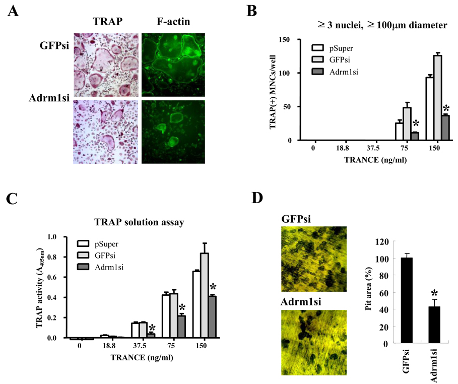
Fig. 3.
Adrm1 expression during osteoclast differentiation. BMMs were infected with retrovirus alone (pSuper), control siRNA (GFPsi), or siRNA specific for Adrm1 (Adrm1si). The infected cells were cultured with 60 ng/ml M-CSF and with the indicated concentrations of RANKL. (A) After 4 days of culture, cells were fixed and stained for TRAP and F-actin. (B) TRAP+ MNCs with more than three nuclei and a diameter larger than 100 µm were counted as osteoclasts. (C) TRAP solution assays were performed as described in the Materials and Methods. (D) Pit formation. BMMs were cultured on dentine slices with M-CSF and RANKL for 4 days. Resorption pits were stained with 0.5% toluidine blue and the pit area was measured using image analysis software. Data are expressed as the mean ± SD and are representative of at least three independent experiments. *, P < 0.05 versus vector controls.

