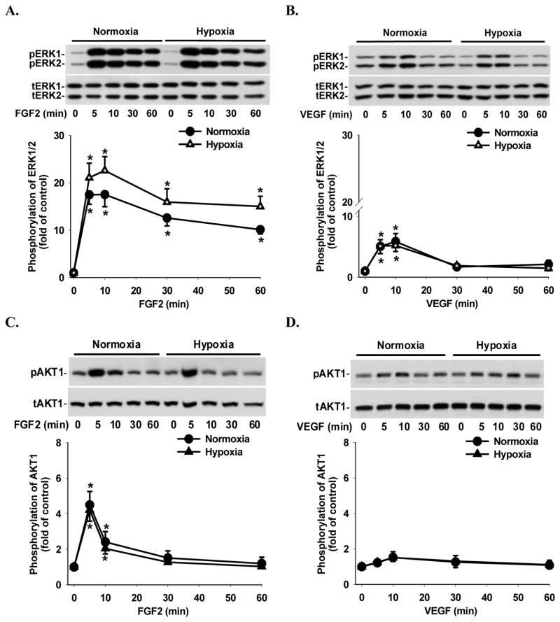
Figure 3.
Effects of hypoxia on FGF2- and VEGF-induced ERK1/2 and AKT1 phosphorylation in HPAE cells. Cells plated in 6 cm culture dishes were cultured under normoxia until reaching 70–80% confluence, followed by normoxic or hypoxic culture for 24 hr and serum starvation for additional 24 hr. Cells were then treated with 10 ng/ml of FGF2 (A and C) or VEGF (B and D) for 0–60 min. Proteins were subjected to Western blot analysis for total ERK1/2 (tERK1/2) and total AKT1 (tAKT1) or phospho-ERK1/2 (pERK1/2) and phospho-AKT1 (pAKT1). Data normalized to tERK1/2 and tAKT1 are expressed as means ± SEM fold of the time 0 controls (n = 6). *differ (p < 0.05) from the corresponding time 0 control.

