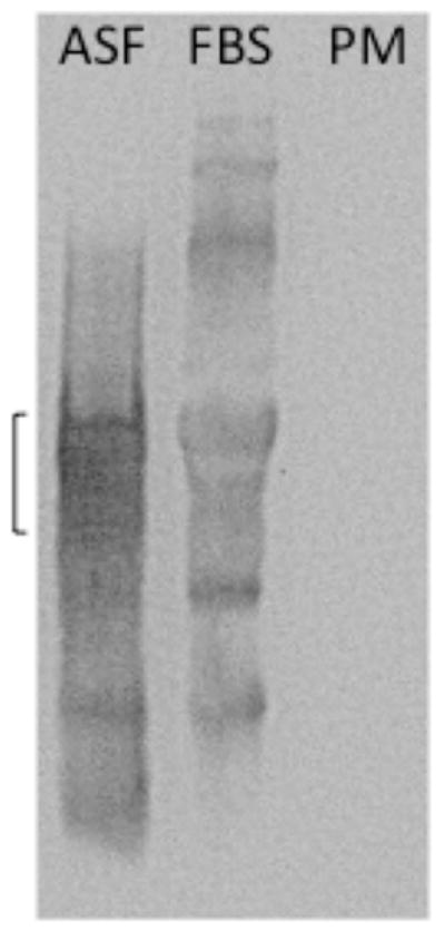
Figure 2.

Absence of detectable levels of ASF in the plasma membrane fraction. ASF (10ug), FBS (20ul) and plama membrane proteins (PM, 5ug) were separated by SDS-PAGE on a 10% gel and transferred to a PVDF membrane. ASF was detected using a sheep anti-bovine ASF IgG polyclonal antibody (1:1000) and a HRP-conjugated donkey anti-sheep IgG secondary antibody (1:2000). Bands were visualized using Super-Signal West Pico Chemiluminescent Substrate and exposure in the SynGene Gnome. The bracket indicates the molecular weight region (~ 61kDa) in which ASF is found [17]. Because ASF is glycosylated it is not found as a tight band.
