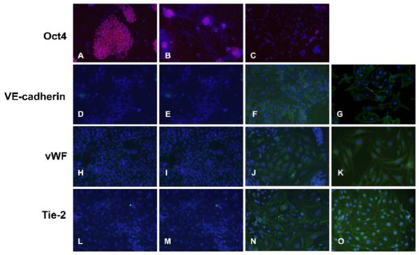
Figure 3. Immunocytochemical analysis of expression of the pluripotency gene Oct4 and EC marker genes VE-cadherin, vWF and Tie2 in response to DNMT inhibition in ESC.

The ESC were exposed to vehicle or 1 μM or 3 μM aza-dC for 24 hours in ESGRO COMPLETE™ basal media lacking LIF. Cultures were returned to basal medium without aza-dC after 24 h of treatment and cells were stained as described in Methods after 15d of treatment. Nuclei are stained in blue by DAPI. Images were captured at 200x magnification. Oct4 :(A) ESC, (B) vehicle-treated ESC, (C) ESC treated with 3 μM aza-dC. VE-cadherin : (D) ESC, (E) vehicle-treated ESC, (F) ESC treated with 3 μM aza-dC, (G) mBEC. vWF : (H) ESC, (I) vehicle-treated ESC, (J) ESC treated with 3 μM aza-dC (K) mBEC. Tie2 : (L) ESC, (M) vehicle-treated ESC (N) ESC treated with 3 μM aza-dC (O) mBEC. mBEC were used as positive controls. The experiment was repeated three times and representative data is presented.
