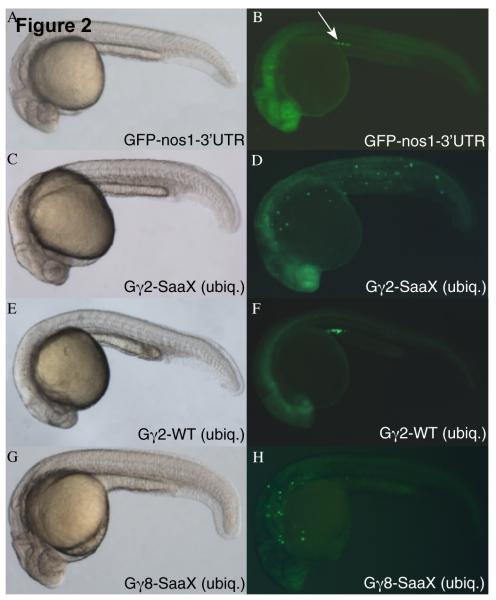
Fig. 2.
PGC migration in embryos overexpressing WT and SaaX mutated Gγ subunits. Bright-field image of an embryo injected with GFP-nos1-3′UTR mRNA [150pg] (A) results in GFP fluorescence restricted to the PGCs located at the anterior of the yolk extension (arrow in B). Bright-field image of an embryo injected with a mix of gng2-SaaX mRNA [20pg] driven ubiquitously (ubiq.) by the Xenopus β-globin 5′UTR & 3′UTR and the PGC driven GFP-nos1-3′ UTR mRNA (C). Fluorescent image of (C) reveals PGC migration defects (D) that are not observed when embryos are injected with gng2-WT mRNA [400pg] (E & F). Bright-field image of an embryo expressing gng8-SaaX [40pg] reveals no obvious alteration in somatic development (G) but fluorescent microscopy of the same embryo indicates PGC migration is perturbed (H). All embryos shown were injected with mRNAs at the 1 – 4 cell stage and subjected to microscopy at 24hpf.

