Fig. 5.
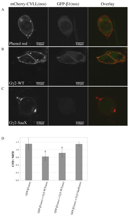
The membrane localization of Gβ1 is lost in the presence of Gγ-SaaX. Confocal images of PGCs migrating in vivo after embryos were injected with the plasma membrane-localized mCherry-CVLL(nos) mRNA (column 1) and GFP-gnb1 mRNA (column 2). Embryos were co-injected with either phenol red as a control (A), gng2-WT mRNA (B), or gng2-SaaX mRNA (C). Ratios of the GFP fluorescence density in the cytosol to the GFP fluorescence density in the membranes of embryos injected with or without Gγ subunits (D) (CFD = cytosol fluorescence density; mfd = membrane fluorescence density; * p < 0.002 when compared to phenol red and gng2-SaaX injected groups). (For interpretation of the references to color in this figure the reader is referred to the web version of this article.)

