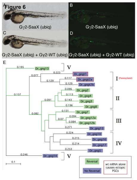
Fig. 6.
Several wildtype G-protein γ subunits have the ability to reverse the Gγ2-SaaX induced disruption of PGC migration. Bright-field microscopy of a larvae ubiquitously expressing gng2-SaaX [20pg] reveals no somatic defects (A) while PGC migration in this same embryo is perturbed (B). Bright-field image of a larva ubiquitously expressing both gng2-WT mRNA [200pg] and gng2-SaaX mRNA [20pg] (C). The fluorescent image of (C) indicates a restoration of proper PGC migration (D). All embryos shown were injected at the 1 – 4 cell stage with mRNAs as described above, together with GFP-nos1-3′UTR mRNA and subjected to microscopy at 48hpf. Phylogenetic analysis of the zebrafish Gγ subunits and their ability to reverse the Gγ2-SaaX induced disruption of PGC migration (E). Method: Neighbor Joining; Best Tree; tie breaking = systematic. Distance: Poisson-correction; Gaps distributed proportionally. The Gγ subunits are divided into five groups according to their secondary structure as previously described [73]. Data was summarized from 2 – 5 experiments with a minimum of 20 embryos scored for the presence of ectopic PGCs at 24hpf. (For interpretation of the colors in this figure the reader is referred to the web version of this article.)

