Fig. 9.

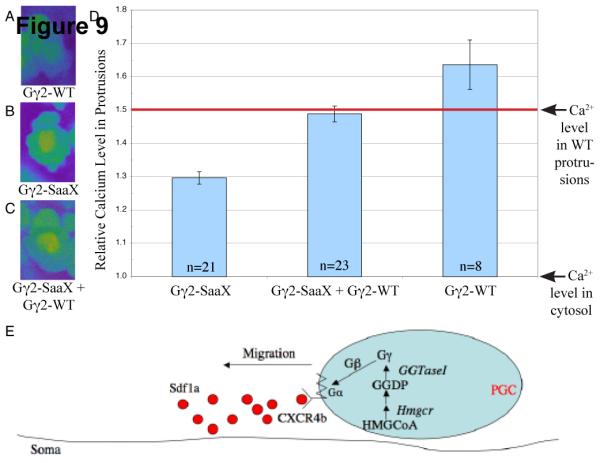
Overexpression of gng2-SaaX attenuates calcium accumulation within the protrusions of PGCs migrating in vivo. Injection of embryos at the 1-cell stage with the PGC-driven gng2-WT mRNA [75pg] results in an increase in the accumulation of calcium in the protrusions of PGCs migrating in vivo (A). PGC-driven gng2-SaaX mRNA [50pg] results in a suppression of the calcium accumulation in these protrusions (B), which is reversed by injection with gng2-WT mRNA [75pg] (C). Calcium levels were quantified with the calcium indicator Oregon green 488 BAPTA-1 [500 μM]. To identify the PGCs, embryos were co-injected with vasa-DsRedex1-nos1 RNA [180 pg] (A-C) and data is summarized in (D). (All PGCs described above were assayed 9-11hpf.) Model of a cell autonomous mechanism for HMGCR’s role in PGC migration (E). Internalization of the extracellular signal Sdf1a is transduced through a GPCR that utilizes heterotrimeric G-Proteins of which the γ subunit is a substrate for geranylgeranylation. (HMGCoA = 3-hydroxy-3-methylglutaryl-Coenzyme A; HMGCR = HMGCoA Reductase; GGDP = geranylgeranyl diphosphate synthase; GGTaseI = geranylgeranyl transferase I) (For interpretation of the colors in this figure the reader is referred to the web version of this article.)
