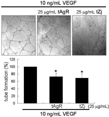
Fig. 4.
Representative images showing tube formation in VEGF-treated endothelial cells plated onto matrigel. After 12 h incubation with 25 µg/mL tAgR and tZj, cells were fixed and images at ×100 magnification were captured. The number of tubes from the images was counted. Multiple five random fields of view were analyzed for the quantitative results. Tube formation results are plotted as means ± SEM (n=4). *P<0.05, compared to VEGF-untreated control.

