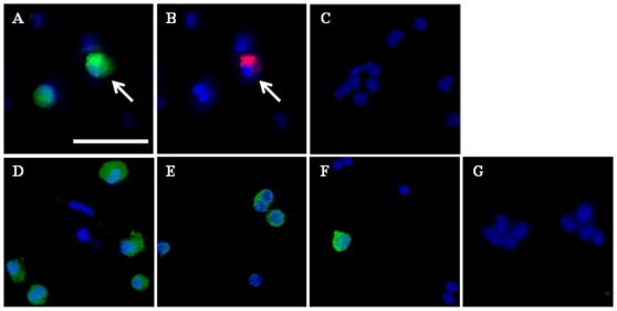
Figure 3. Detection of germ markers in PGC-like cells.
Immunolocalization of A) OCT4, B) VASA, D) STELLA, E) c-KIT, and F) DAZL in D30 PGC-like cells. White arrows indicate cells co-staining for OCT4 and VASA (A and B). Controls in which cells were probed with secondary antibody alone (either anti-rabbit or anti-mouse-FITC/PE) were negative (C and G, respectively). Counter-staining with Hoechst was conducted to detect nuclei. Panels are shown at 400X magnification (bar = 50 µm).

