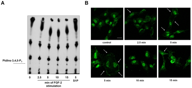
Figure 3. FGF-2 activates PI3K in HUVEC.
(A) TLC analysis of phospholipids extracted from [32P]-labelled HUVEC stimulated with 100 ng/ml FGF-2 for the indicated times or 1 µM sphingosine-1-phosphate (S1P) for 5 min. Arrow indicates the PtdIns-3,4,5-P3 spot. (B) HUVEC grown on glass coverslips were incubated overnight in M199+0.5%FBS before stimulation with 100 ng/ml FGF-2 for the indicated times. Coverslips were then incubated with an anti PtdIns-3,4,5-P3 antibody and analyzed by confocal microscopy. Arrows indicate the plasma membrane localization of the phosphoinositide. Bar = 10 µm.

