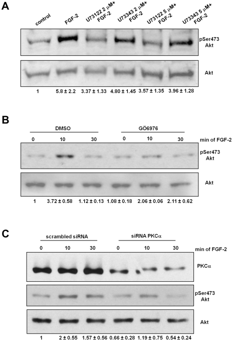
Figure 6. PLCγ1-dependent PKCα activation mediates FGF-2-induced Akt activation.
HUVEC were incubated overnight in M199+0.5%FBS and then treated with the indicated concentrations of U73122 or U73343 for 30 min (A) or 10 µM of the conventional PKCs inhibitor Gö6976 (B) before stimulation with 100 ng/ml FGF-2 for 10 min. (C) HUVEC were transfected with scrambled siRNA or siRNAs targeting PKCα. After 24 h cells were serum deprived overnight and then stimulated with 100 ng/ml FGF-2 for the indicated times. Phosphorylation of Akt at Ser473 was assessed by using a specific antibody. Membranes were then stripped and re-probed with an anti Akt. Blots are representative of 7 (A) and 3 (B,C) independent experiments. In all cases, quantitative analysis of the band intensities was performed. Data represent values of pSer473 Akt/Akt expressed as fold increase over control.

