
Figure 2.
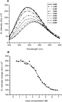
(A) Fluorescence spectra of HCoV-OC43 N protein in Tris-HCl supplemented with different concentrations of urea. The protein concentration was 1 μM and the buffer consisted of 50 mM Tris-HCl (pH 7.3), 150 mM NaCl, and 0.1% CHAPS. (B) Urea-induced unfolding of HCoV-OC43 N protein monitored by fluorescence emission at 340 nm. The protein concentration was 1 μM and the buffer consisted of 50 mM Tris-HCl (pH 7.3), 150 mM NaCl, and 0.1% CHAPS.