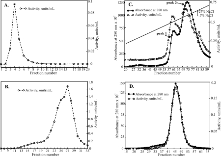
Figure 2.

Steps involved in the purification of hEpman: Graphs show α-mannosidase activity, units/mL (open circles, dashed line) and absorbance at 280 nm (filled circles, solid line). (A) Cibacron Blue F3GA step eluted with 500 mM NaCl. Fractions 1-10 were pooled. (B) Pooled fractions bound to cobalt chelating Sepharose and eluted with pH 5.0 acetate buffer. Fractions 21–30 were pooled for the next step (C) Cation exchange chromatography on SP-HiTrap with an applied 0.05–0.27 M salt gradient. Fractions from peaks 1 and 2 were pooled separately (D) Final purification step of peak 2 on a Superdex 200 16/60.
