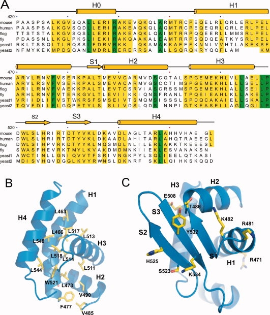
Figure 3.

(A) Primary and secondary structure of mCdt1C showing identity with orthologues from human, frog, fly, worm, and yeast (1: Schizosaccharomyces pombe and 2: Saccharomyces cerevisiae). Highly conserved residues are colored with yellow bar and absolutely conserved residues are colored with green. (B) Conserved hydrophobic residues in mCdt1C are shown in yellow stick. (C) Conserved exposed residues in the mCdt1C core are clustered in three β-strands.
