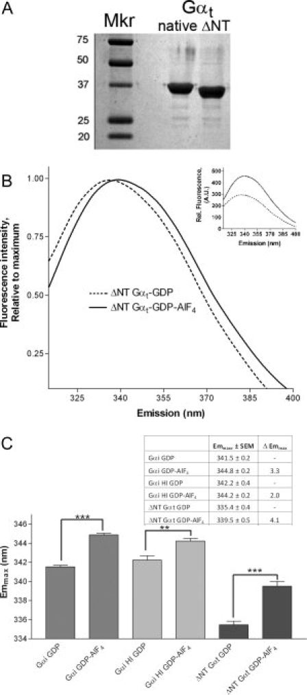
Figure 3.

Trp fluorescence of unmyristoylated proteins. (A) Myristoylated Gαt (native) is enzymatically cleaved at the N-terminus to yield the unmyristoylated ΔNT Gαt protein used in structure determination.6 (B) Emission of 500 nM ΔNT-Gαt protein was scanned as above before (dotted line) and after (solid) activation with AlF4 as in Figure 1. Control, inset: ΔNT-Gαt demonstrated ≥40% increase in Trp fluorescence upon AlF4 activation. (C) Summary of emmax before and after activation for indicated Gα proteins (n ≥ 3, ±SEM; statistically significant changes noted by asterisk(s), ***P < 0.001, **P < 0.01). Dark gray bars, far left, Gαi; light gray, center, Gαi HI; black, far right, ΔNTGαt.
