
Figure 1.
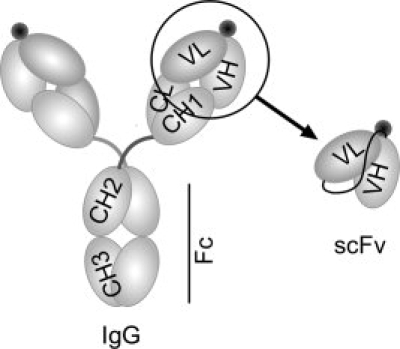
The structural representation of an intact IgG and the scFv antibody. To convert the anti-METH IgG6H4 binding region to anti-METH scFv6H4, the variable heavy (VH) and variable light (VL) regions were amplified and joined by PCR. The antigen binding sites are represented by black circles near the VH and VL domains.