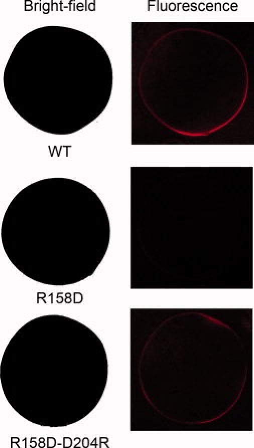
Figure 7.

Bright-field and fluorescence images obtained with oocytes labeled with anti-GABAC ρ1 antibody. Top row: Oocyte expressing the wild-type GABAC ρ1 receptor. Middle row: Oocyte expressing the R158D mutant ρ1 GABAC. Bottom row: Oocyte expressing the double mutant R158D–D204R ρ1 GABAC. These results show that the partially degraded R158D mutant did not express on the surface of the oocyte, while the surface expression was rescued by the additional mutation at Position 204.
