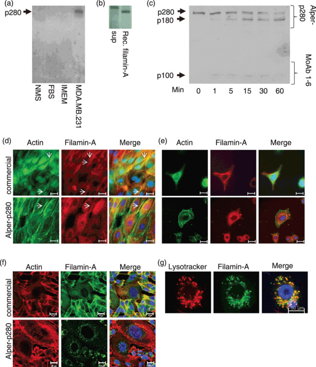
Figure 1.

Characterization of p280. Alper‐p280 was used to western blot normal mouse serum (NMS), fetal bovine sera (FBS), concentrated Iscove's modified essential medium (IMEM), and culture medium conditioned by MDA.MB.231 cells (a). MDA.MB.231 conditioned media (sup) and recombinant filamin‐A were resolved by SDS‐PAGE followed by electrotransfer to nitrocellulose. Western blots were then probed using Alper‐p280 (b). MDA.MB.231 culture supernatant was subjected to calpain cleavage for the indicated times. Proteins were resolved by SDS‐PAGE, transferred to nitrocellulose, and then blotted using Alper‐p280 (c). Normal human mammary epithelial cells (HMEC) were fixed, permeabilized, and stained using rhodamine‐conjugated phalloidin (column 1, green), and filamin‐A using either a commercially available anti‐filamin‐A antibody (top middle panel, red) or Alper‐p280 (bottom middle panel, red). Far right hand panels are merged images. Scale bars = 20 µm (d). SKBR3 and MDA.MB.231 breast cancer cells were stained as described in (d). Scale bars = 20 µm (e,f). Lysosomes of MDA.MB.231 cells were labeled using lysotracker, fixed, and then permeabilized. Cells were stained with Alper‐p280 followed by Alexa488‐conjugated anti‐mouse secondary antibody. Scale bars = 50 µm (g). All data are representative of three independent experiments.
