Figure 1.
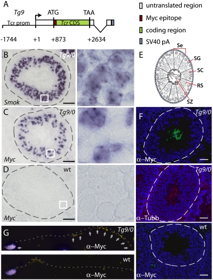
Tcr transcripts escape the general mechanism of product sharing between syncytial sperm cells and are translated at late stages of spermiogenesis. (A) Schematic drawing of the transgene construct Tg9 comprising the Tcr promoter (Tcr prom), untranslated region, and Myc epitope-tagged Tcr coding region (Tcr CDS), followed by the simian virus polyadenylation signal (SV40pA). (B–D) Cryosections of Tg9/0 (B,C) or wild-type (D) testes hybridized in situ with digoxygenin (DIG)-labeled Smok1-specific (B) or Myc epitope-specific (C,D) probes, showing expression of endogenous Smok1 transcripts in all round spermatids (B), whereas Tg9 transcripts are confined to a subpopulation of round spermatids (C) and not detected in the control (D). Dashed lines indicate the outlines of seminiferous tubules, and white boxes indicate subregions shown at higher magnification on the right. Bar, 100 μm. (E) Schematic view of a seminiferous tubule. (Se) Sertoli cells; (SG) spermatogonia; (SC) spermatocytes; (RS) round spermatids; (SZ) spermatozoa. (F, top panel) Immunofluorescent detection of Myc-Tcr protein using an anti-Myc antibody (α-Myc) on testis cryosections of a Tg9/0 male reveals Tcr protein in the lumen of seminiferous tubules. (Middle panel) β-Tubulin (α-Tubb), a major component of axonemes, indicating flagella, localizes to the same region. (G) In epididymal spermatozoa, Myc-Tcr localizes primarily to the principal piece and nucleus (yellow arrows; the flagellum is indicated by a dashed line). (F,G, bottom panel) Wild-type controls show some nonspecific primary antibody reaction. Nuclei are visualized by DAPI staining (blue). Bar, 50 μm.

