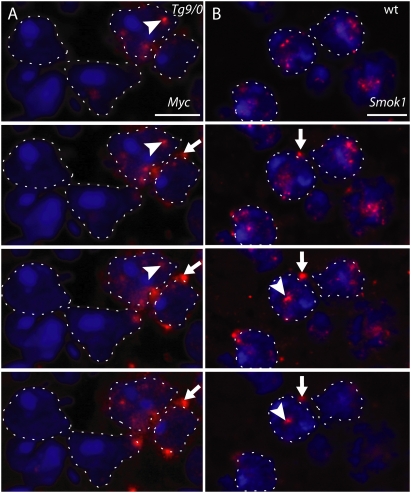
Figure 2.
Tcr and Smok1 transcripts occur mainly in nuclear and perinuclear aggregates. Serial optical sections (∼1-μm depth) of testicular cryosections obtained by confocal microscopy visualizing Tg9 (A) or Smok1 (B) transcripts (red) detected by fluorescent in situ hybridization in round spermatids. Selected examples of nuclear (arrowhead) and perinuclear (arrow) RNA aggregates are indicated. Tg9 transcripts were detected on sections of Tg9/0-derived testes (shown in A) with a DIG-labeled Myc epitope-specific probe, and Smok1 transcripts were detected on sections of wild-type testes with a Smok1-specific probe. Dashed lines denote the nuclear outlines of round spermatids visualized by DAPI staining (blue), and bright blue structures within the nuclei represent nucleoli. Bar, 6.5 μm.

