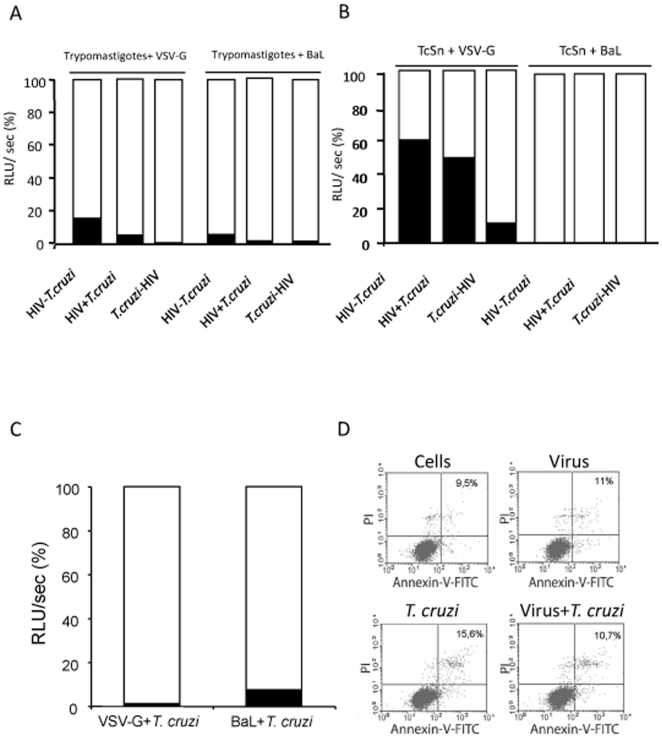
Figure 3. Inhibition of pseudotyped viruses replication by T. cruzi.
MDM were infected with both VSV-G or BaL pseudotyped viruses in the presence of T. cruzi blood trypomastigotes (A) or trypomastigotes free-supernatant (TcSn) (B) in three different schemes: HIV 24 h after T. cruzi (T. cruzi-HIV), HIV 24 h before T. cruzi (HIV-T. cruzi) or HIV at the same time as T. cruzi (HIV+T. cruzi), where T. cruzi indicates either trypomastigotes of TcSn. Luciferase activity was measured from cell lysates 4 days post-viral infection. Results are expressed as relative light units per second (RLU/sec), presented as a percentage relative to the control (100%), where the histogram in white corresponds to the percentage of infection with the respective control virus and the histogram in black corresponds to the percentage of infection in the presence of the parasite. (C) MDM were also infected with pseudotyped viruses and cell-derived trypomastigotes, and luciferase activity was evaluated. (D) Cell viability was evaluated by flow cytometry at day 4 p.i. in co-infected cells with blood trypomastigotes and controls stained with PI and Annexin-V-FITC. During the analysis 20000 events were acquired and the analysis includes all the ungated cells; percentages of PI and Annexin-V positive cells are indicated. Results are representative of 3 independent experiments performed with cells from different donors.

